Visual Field Examination
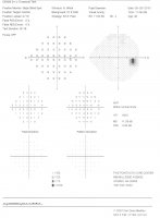 By examining a person's peripheral vision, a visual field test can help us diagnose many different conditions ranging from ocular migraines to macular degeneration to glaucoma and even brain tumors. A visual field test consists of a series of lights flashing at varying intensities in random positions around the patient's peripheral vision, with some lights occasionally flashing in front. While looking straight ahead, the patient depresses a button every time they perceive a flash of light. Once both eyes have been tested, the pattern that results helps us determine potentially damaged areas of the retina, damage to the optic nerve or even the possibility of a greater problem such as a brain tumor. Should there be such a finding, an accurate visual field test can even help narrow the search for a potential tumor as it will often indicate a fairly specific locale based on the pattern of the results.
A visual field test evaluates the performance of the visual pathway. The visual pathway is the path the nerves from the eye take through the brain to get to the back of the brain where all the visual information from the eyes is processed. Since this pathway is so long, many problems in the brain can affect it and therefore affect vision.
Some of the diseases that affect the visual fields are:
- Glaucoma
- Optic nerve diseases and tumors
- Stroke
- Multiple Sclerosis
- Brain tumors or aneurysms
- Pituitary tumors
- Brain injury/trauma
Optical Coherence Tomographer (OCT)
 The Optical Coherence Tomographer or OCT uses special light to scan the optic nerve and retina, creating detailed images that can help doctors manage glaucoma, other optic nerve diseases and retinal diseases like macular degeneration and macular edema (swelling).
OCT is one of the latest high-tech diagnostic tools in the defense against glaucoma.Glaucoma, usually indicated by abnormally high pressure inside the eye, can develop into blindness. OCT can help us determine a patient's potential to develop glaucoma before tissue damage occurs, allowing us to consider treatment options long before other tests would have even diagnosed the condition. The sooner glaucoma is detected, the better the chance of preserving vision.
When OCT scans are performed on the retina, the images produced give a cross-section of the macula in such detail that the different layers of the retina can be distinguished which allows better diagnosis of retinal diseases. With this diagnostic tool treatment for macular degeneration and retinal swelling from diabetes can be monitored, enabling our Athens eye doctors to assess whether or not more treatments are necessary.
Digital Retinal Photography
 Digital retinal photographs are pictures of the inside of your eye taken by a special digital retinal camera. The photographs are used by the doctor to monitor and identify any changes in the health of your retina and the optic nerve. The retina and optic nerve need to be healthy in order for you to see clearly.
Many systemic diseases can affect the retina, including: diabetes, hypertension, arteriosclerosis, high cholesterol, and AIDS. The photographs show the macula, the part of the retina that is responsible for your detailed, sharp vision. We can monitor macular degeneration and other diseases of the macula with these photographs. The pictures also show the optic nerve, which carries all of the information from your eye to your brain. Damage to the optic nerve can cause glaucoma and loss of sight.
Retinal photographs are important for documenting retinal problems as well as for monitoring your retinal and optic nerve health. Baseline retinal photos can help our Athens eye care experts to determine if any changes have occurred in your eye health when you have your comprehensive eye examinations.
Corneal Topography
 The cornea is the highly curved clear window on the front of the eye, right above the iris, the colored part of the eye. The cornea helps focus light on the retina and therefore plays a significant role in how well you see. As the curvature of this clear structure changes, both the way your contact lens fits and your prescription, can change. Understanding the curvature of the cornea is necessary to properly fit and prescribe contact lenses or spectacles.
The corneal topographer is a highly sophisticated computer that "maps" the front surface of the eye to show how curved it is. Abnormalities in the curvature of the cornea can help our Athens optometrist diagnose corneal diseases and determine the best fit for specialty contact lenses. This information can also help your doctor determine if refractive surgery is appropriate for you. This computer can also help monitor subtle changes on your cornea over time and can also be useful when determining the extent of damage in the case of an eye trauma.
The corneal topography analysis uses colors to create a model of the cornea. The map colors indicate how the cornea flattens and steepens (the valleys and hills) over its entire surface. The corneal topographer analyzes the shape of the cornea several different ways to help your Athens optometrist evaluate its health.
Pachymetry
 Pachymetry is the measurement of the thickness of the cornea. This special medical device is used to help with the diagnosis of glaucoma. One of the causes of glaucoma is having too much pressure inside the eye, which damages the optic nerve and causes loss of sight. When we measure the pressure inside the eye with tonometry, the readings can be affected by how thick or thin the cornea is; thick corneas can make the pressure measured artificially high and thin corneas measure it artificially low. Having thinner corneas is a risk factor for glaucoma, making pachymetry an important part of a glaucoma work-up.
Gonioscopy
 Gonioscopy is used to visualize the anterior chamber angle of the eye, which is the front part of the eye between the cornea and the iris. This structure cannot be seen unless a special lens, called a gonioscopy lens, is placed on the eye. The doctor then uses the mirrors in the gonioscopy lens to assess the health of this portion of the eye.
Gonioscopy is most often done if glaucoma is suspected. Before a diagnosis of glaucoma can be made, gonioscopy is done to determine which type of glaucoma is present. Glaucoma is a group of eye disorders in which a specific pattern of damage occurs to the optic nerve, resulting in loss of eyesight. Gonioscopy is done to determine whether the area in the eye where fluid drains out of the eye (called the anterior chamber angle) is damaged, blocked, or clogged. It is often done as part of a complete glaucoma exam. It may also be done during a routine eye exam, depending on your age and whether you are at high risk for developing glaucoma.
Visit our Athens optometrist for emergency eye care today!